Researchers developed AI models that use a single facial photo to estimate age and assess mortality risk in cancer patients.
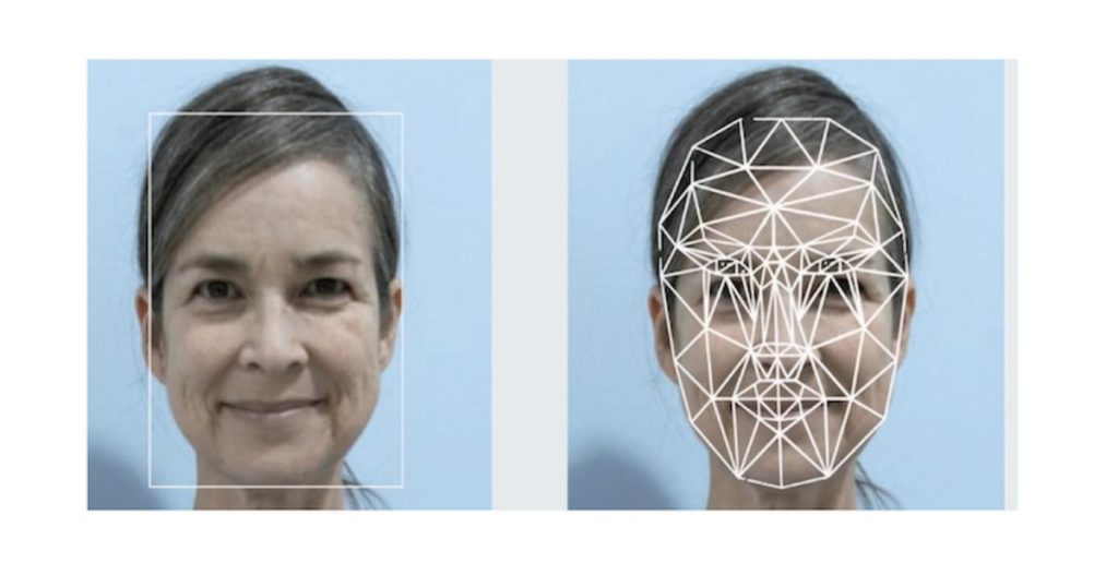
What You Need to Know
- FAHR-Face, a system trained on more than 40 million facial images, was used to develop FAHR-FaceAge, which estimates cancer patients’ facial age from a single photograph. A facial age at least 5 years older than a patient’s chronological age was associated with a 21% higher risk of death compared with the reference group.
- Some facial regions analyzed by the models resemble signs described by Hippocrates about 2,400 years ago, including a pinched nose, sunken eyes, and hollow temples — a similarity highlighted by the study’s authors. The models were not trained on ancient texts.
- In the highest-risk group identified by FAHR-FaceSurvival, 52.5% of the 122 patients died within 90 days.
A team at Mass General Brigham, a Boston-based medical and research system in Massachusetts, developed AI models that estimate a cancer patient’s facial age and risk of death from a single facial photograph. The findings were published on October 19, 2026, in The Lancet Digital Health.
How the AI Models Work
The models are based on FAHR-Face, a system trained on more than 40 million facial images. Two tools were developed from it:
- FAHR-FaceAge: Estimates how old a person looks. The gap between estimated facial age and chronological age was linked to risk of death.
- FAHR-FaceSurvival: Generates a survival score from a facial photograph, with higher scores indicating a greater risk of death.
Each algorithm focuses on different areas of the face:
- FAHR-FaceAge: Homes in on the nasolabial fold, the crease running from the sides of the nose to the corners of the mouth, as well as the forehead above the eyes and the temples.
- FAHR-FaceSurvival: Mainly analyzes the area under the eyes and the bridge of the nose, between the eyes.
According to the authors, this is the first demonstration that AI can estimate cancer survival directly from a simple photograph.
An Echo of Hippocrates
Some of the facial regions the models flag resemble signs described by Hippocrates, considered the father of medicine, around 400 BC: “a sharp nose, hollow eyes, sunken temples.”
The models were not trained on the ancient texts. They were developed using facial images and adapted to estimate age or assess survival, depending on the task. Radiation oncologist Raymond Mak called the similarity striking, describing it as “Hippocrates echoing across the millennia.”
Key Findings
Among cancer patients treated with radiation therapy in the US, with a median follow-up of 4.3 years, researchers found associations between estimated facial age and risk of death:
- Facial age at least 5 years older than chronological age: A 21% higher risk of death than the reference group in this analysis.
- Facial age at least 5 years younger than chronological age: A 17% lower risk of death, based on the specific comparison used in the study.
Deaths Within 90 Days of Assessment
- FAHR-FaceSurvival: In the highest-risk group, 52.5% of 122 patients died. In the lowest-risk group, about 2% died (20 of 975 patients).
- FAHR-FaceAge: Among patients whose estimated facial age was more than 20 years above their chronological age, 21.3% died (of 1,060 patients). Among those whose estimated facial age was at least 10 years below their chronological age, 6.4% died (of 4,195 patients).
The study also found statistical associations between estimated facial aging and factors such as heavy alcohol use, drug use, and smoking. That does not show that these factors alone explain the models’ estimates.
FAHR-FaceAge produced consistent results despite changes in lighting, angle, facial expression, and makeup. Most cosmetic procedures tested did not significantly affect performance, though a small difference was observed after rhinoplasty.
Potential Use
In the future, facial analysis could complement assessments of a patient’s physical condition before intensive treatments such as radiation therapy, surgery, and systemic therapy.
According to Mak, the goal is to make this assessment more quantitative, consistent, and scalable, helping support personalized decisions without replacing clinical judgment. The researchers also plan to explore whether the technology could provide useful health insights for people without cancer, a hypothesis that requires further study.
Expert Caveats
- Wilbur Lam, Emory University: The associations appear robust, but clinical utility must be proven in prospective studies. Performance also needs to be evaluated across different skin tones, age groups, and patient populations.
- Miroslaw Bober, University of Surrey: Prediction is not explanation. The AI estimates age from facial appearance without directly measuring the biological processes of aging. The study also did not show whether the predictions add clinically useful information to conventional medical assessments.
AI facial analysis could become a complementary tool for evaluating cancer patients. However, the technology is not yet ready to be used as a standalone tool for clinical decision-making.